

Researchers at Swansea College have developed a novel strategy to testing biomaterials for bone regeneration, providing each velocity and moral refinement.
Revealed in Bioactive Supplies journal, and led by Dr. Zhidao Xia, a workforce from Swansea’s Medical College and School of Science and Engineering has launched a murine tibial periosteal ossification mannequin that eliminates the necessity for conventional strategies involving fractures or essential sized defects.
In doing so, they considerably scale back animal struggling whereas enabling biomaterial analysis in as little as 14 days, with cortical bone transforming accomplished by 28 days.
Dr. Xia defined, “Our invention bridges the hole between artificial substitutes and donor bone. We’ve proven that it’s doable to create a cloth that’s secure, efficient, and scalable to satisfy world demand. This might finish the reliance on donor bone and deal with the moral and provide points in bone grafting.”
Alongside Swansea, a number of establishments worldwide contributed to this venture. These embody the likes of Huazhong College of Science and Know-how, and Xiangyang Central Hospital from China, Johns Hopkins College College of Medication and the College of Rochester from USA, McGill College from Canada, Oxford Devices NanoAnalysis, The Open College, the College of Oxford, and the College of Sheffield from UK.
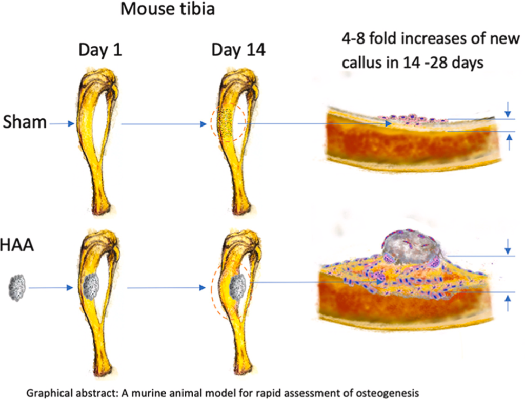
Periosteal mannequin improves bone restore testing
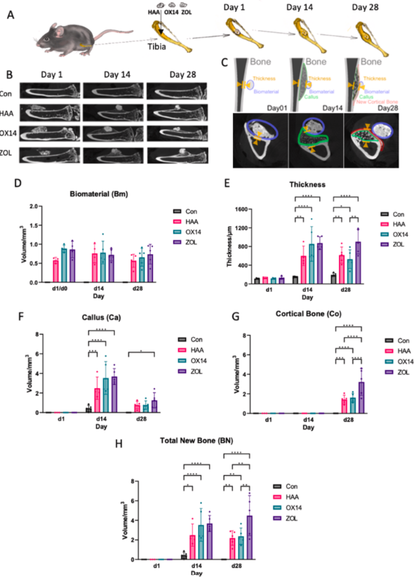
Central to the mannequin’s success is its concentrate on the tibial periosteum, the place osteogenic cells naturally drive bone restore. Implanting biomaterial scaffolds between the tibia and surrounding muscle tissue has yielded notable outcomes, together with trabecular bone progress inside two weeks.
By the fourth week, this bone remodels into a brand new cortical layer. These findings, measured via micro computed tomography and histological evaluation, revealed an distinctive eightfold enhance in tibial thickness in comparison with management teams.
One of many supplies examined on this mannequin is a hydroxyapatite aragonite (HAA) scaffold, a mix of hydroxyapatite extensively utilized in bone grafts and calcium carbonate, which reinforces biodegradation and helps bone progress.
The scaffold progressively breaks down over six to 12 months, creating house for brand spanking new bone tissue to develop. Researchers additionally experimented with zoledronate, a medical bisphosphonate, discovering that its addition considerably elevated cortical bone formation, demonstrating its potential in enhancing biomaterial efficiency.
In contrast to ectopic strategies the place bone is generated in unnatural places, this strategy maintains the fabric’s direct contact with each bone and muscle. This proximity replicates the advanced environments sometimes encountered in orthopaedic purposes, making the findings extremely related for medical eventualities.
Validation of the mannequin throughout species together with rats and minipigs confirmed constant outcomes. The HAA scaffold not solely facilitated bone regeneration but additionally degraded totally inside six months, in keeping with the researchers.
Moral refinements and future instructions
A notable benefit of the mannequin lies in its streamlined testing course of. Eliminating the necessity for giant bone defects or fractures considerably reduces the invasiveness and period of experiments.
Whereas the method is especially fitted to early stage screening of biomaterials, bigger animal trials and conventional fracture fashions are nonetheless required for regulatory approval and testing in advanced defect eventualities.
Additional investigations goal to make clear the function of particular cells corresponding to periosteal stem cells within the noticed fast bone formation. Enhancements to micro computed tomography software program are additionally wanted to higher analyze biomaterial efficiency, as present instruments lack precision for evaluating these interactions.
Regardless of these challenges, the analysis highlights calcium carbonate’s pivotal function in accelerating bone progress. Its quicker dissolution in comparison with hydroxyapatite allows bone cells to infiltrate the scaffold extra effectively, though the precise mechanisms warrant extra examine.
Importantly, this mannequin aligns with moral analysis ideas by considerably lowering animal struggling. The absence of induced fractures or massive bone defects minimizes hurt, providing a refined and humane methodology for early-stage biomaterial screening.
Whereas conventional fracture fashions and enormous animal trials stay important for regulatory approval, this strategy might streamline preclinical testing and scale back the variety of animals required.
Different bone regeneration strategies
Globally, analysis teams have explored various approaches, every providing distinctive benefits in tackling the challenges of bone restore.
Two years in the past, researchers from Carnegie Mellon College and the College of Connecticut developed 3D printed calcium phosphate graphene (CaPG) scaffolds for future bone regeneration purposes.
Examined in vitro and in vivo, the scaffolds demonstrated osteogenic potential by supporting stem cell differentiation and regenerating bone in animal fashions. Utilizing a Direct Ink Writing methodology, the CaPG scaffolds had been discovered to biodegrade and resorb in vivo, selling tissue regeneration with out adversarial results on very important organs.
Revealed in Nature, this examine highlights CaPG’s potential as an economical, customizable, and resorbable various to conventional bone grafts.
Again in 2017, researchers from Tianjin College, the Chinese language Academy of Sciences, and the College of Hong Kong developed a 3D printable hydrogel-nanoclay composite designed to help bone cell progress.
Revealed in ACS Biomaterials Science & Engineering, this materials addresses bone defects brought on by trauma, deformities, or tumor removing. In checks, the fabric promoted bone regeneration in tibia defects of stay rats, considerably outperforming placebo therapies over eight weeks.
Providing structural help and diet, the scaffold mimicked the extracellular matrix and had a possible to supply precision and individualized restore of load-bearing bone defects.
What 3D printing tendencies do the business leaders anticipate this yr?
What does the Way forward for 3D printing maintain for the following 10 years?
To remain updated with the most recent 3D printing information, don’t overlook to subscribe to the 3D Printing Business e-newsletter or observe us on Twitter, or like our web page on Fb.
When you’re right here, why not subscribe to our Youtube channel? That includes dialogue, debriefs, video shorts, and webinar replays.
Featured picture exhibits a murine animal mannequin for fast evaluation of osteogenesis. Picture by way of Swansea College.